
Microscopy is an ideal tool for understanding differences in particle size and shape. Optical and scanning electron microscopy can be used for simple visual examinations and for more involved image analysis techniques. Chemical imaging using the SEM, IR, Raman, and fluorescence microscopy along with image analysis is an excellent tool for understanding tablets and other dosage forms.
RG12525 Shape Difference by Form
At its simplest, the microscope can show differences in particle shape between different solid-state forms. In this case, the difference in shape provided the means to determine that Form 1 is the stable form at ambient conditions and that the enantiotropic transition temperature is 74 C. (see Solid State Analysis for more information)


Example 2 Fiberglass Insulation
One key factor in the insulating properties of fiberglass is the size of the fibers. Microscopy provides an excellent tool for determining fiber diameter among other properties. These values are helpful in product development.

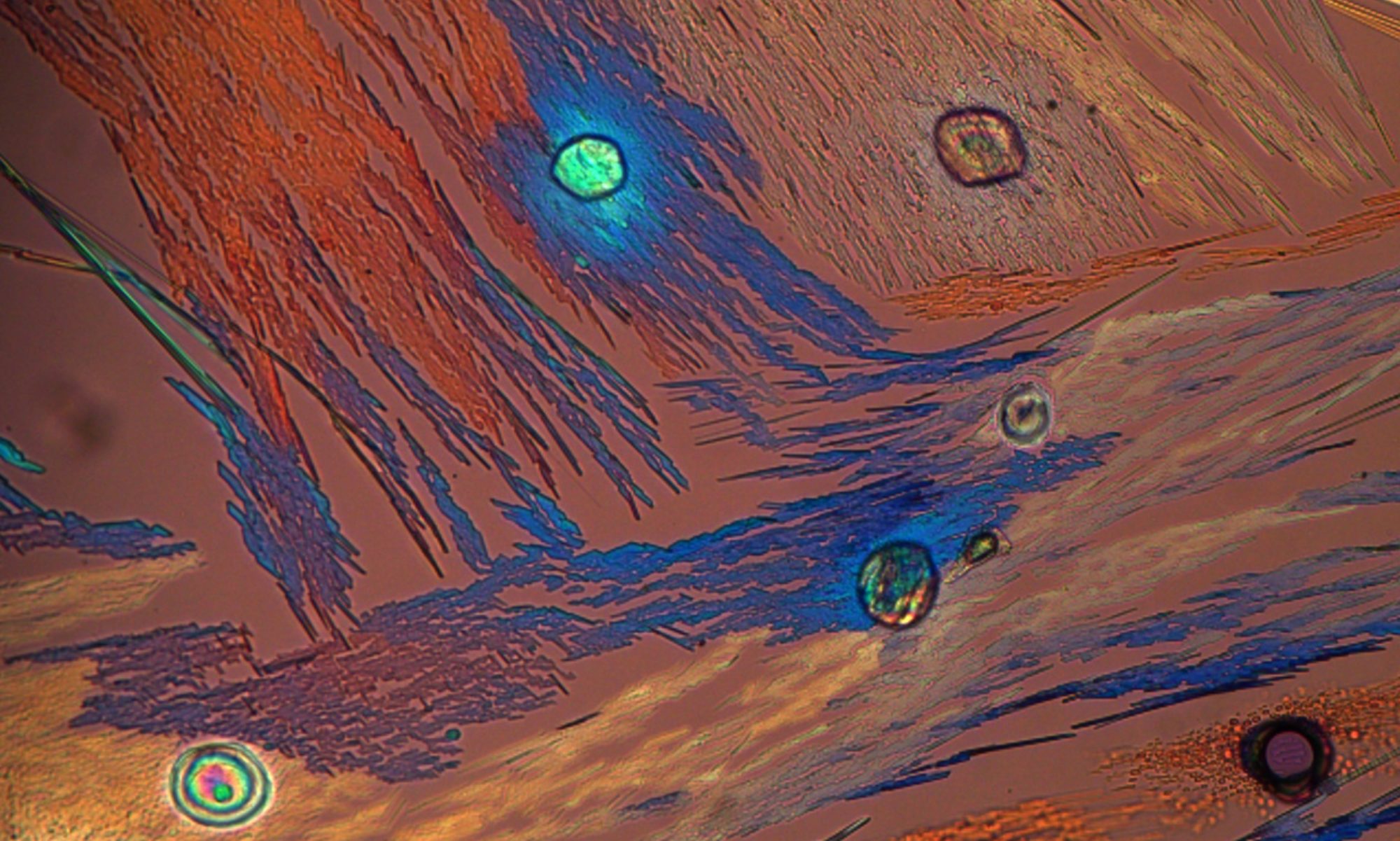
Example 3 Form Conversion in Tablets
Form conversion was occurring in tablets on stability. The conversion process was difficult to measure. With fluorescence microscopy, it was possible to monitor the conversion process and to aid in determining ways to prevent the conversion. (My thanks to Mark Strohmeier for these images).

Summary
There are a host of different ways in which the analysis of particle size and shape can be useful in product development. The microscope provides a direct measurement of both particle size and shape.